Gallbladder Polyps

The Gallbladder and Liver


Gallbladder polyps at a glance
Most gallbladder polyps are benign. Management depends mainly on size, morphology, growth and specific cancer risk factors. Polyps 10 mm or larger usually require surgery. Polyps 6-9 mm may require ultrasound follow-up or surgery depending on risk factors. Very small polyps generally require no follow-up when risk factors are absent.
The gallbladder
The gallbladder is a small pear-shaped organ beneath the liver. It stores and concentrates bile, which is released into the intestine to help digestion.
The polyp
Gallbladder polyps are growths or polyp-like projections from the inner gallbladder wall. They may be flat or attached by a stalk.
They are commonly detected incidentally during abdominal ultrasound. Most are benign (non-cancerous) pseudopolyps, particularly cholesterol polyps. A small proportion are true polyps and could be malignant (cancerous) or have the potential to become malignant. The likelihood of cancer generally increases with size and suspicious morphology.
Causes and risk factors for the formation of gallbladder polyp
The cause depends on the type of lesion. Gallbladder polyps have been associated with cholesterol or salt deposition in bile, gallstones, familial polyposis, Gardner syndrome, Peutz-Jeghers syndrome and hepatitis B.
Classification (types) of gallbladder polyps
Gallbladder polyps are classified into pseudopolyps and true polyps.
Pseudopolyps
The majority (95%) of polyps in the gallbladder are pseudopolyps. They do not have any risk of cancer and generally do not require treatment.
- Cholesterol polyps: The commonest type, caused by cholesterol deposition and often multiple.
- Adenomyomatosis: Benign thickening of the gallbladder wall that may produce a polyp-like appearance.
- Inflammatory polyps: Uncommon lesions associated with gallstones and chronic inflammation.
True polyps
True polyps are less common. They include:
- Adenomas: Benign neoplastic polyps with potential for malignant change.
- Adenocarcinoma: Gallbladder cancer presenting as a polypoid lesion.
Adherent sludge or stones can sometimes mimic a polyp on ultrasound.
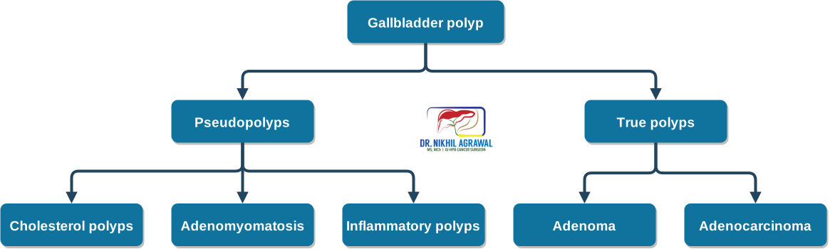
Classification of gallbladder polyp
Symptoms of gallbladder polyps
Most gallbladder polyps cause no symptoms and are discovered incidentally during ultrasound. Some patients have upper abdominal pain, nausea, bloating or food-related discomfort, but these symptoms are nonspecific and may have another cause.
Complications of gallbladder polyp
The principal clinical concern is whether a polyp is noncancerous or cancerous. Risk is low for most small polyps but increases with larger size and suspicious features such as sessile morphology or focal gallbladder-wall thickening. Polyps should therefore be assessed according to size, appearance and patient risk factors.
Diagnosis of gallbladder polyp
Ultrasound (USG): An ultrasound scan uses high-frequency sound waves to create an image of the inside of the body. This is a basic test to see the liver, gallbladder and bile ducts. Most gallbladder polyps are identified on USG. This is also the main modality for following up these polyps when required.
Additional imaging: Repeat ultrasound, Doppler, contrast-enhanced ultrasound, endoscopic ultrasound or MRI may be considered when the finding is indeterminate and better characterization is required.
CT, MRI or PET-CT: These are more useful when cancer is suspected and assessment of disease extent is required.
Risk factors for cancer in gallbladder polyp
Differentiating between cancerous and noncancerous polyps is important. A noncancerous polyp can be observed, while those at risk of cancer are operated upon. One of the most important criteria for this is size.
Size of polyp
A size larger than 10 mm is the most reliable indicator of cancer in a polyp and warrants gallbladder removal surgery (cholecystectomy). A less than 10 mm polyp has a lower chance of harbouring cancer. However, in a study, 10% of cancerous gallbladder polyp were between 5 to 10 mm. Polyps that are less than 5 mm have an extremely low risk of cancer.
Other risk factors for cancer in a polyp
- Age over 60 years
- Asian ethnicity
- Primary sclerosing cholangitis (40-60% polyps in patients with PSC were malignant)
- Flat or sessile polyps
- Focal gallbladder-wall thickening over 4 mm
Treatment of gallbladder polyp
Treatment of gallbladder polyps depends on the risk of cancer in the polyp. Surgical removal of all is not appropriate as the majority of the polyps are pseudopolyps with no risk of cancer.
Those who are at risk of cancer undergo surgery. These include polyps measuring 10 mm or more and may be considered for 6-9 mm polyps when specified risk factors are present. Surgery may also be considered for symptoms attributable to the gallbladder after another cause has been excluded. In those with very low or no risk of cancer, the polyp is only monitored with ultrasound.

Treatment algorithm of gallbladder polyp
Follow-up
Follow-up recommendations vary between societies. Under the updated European joint guideline:
- Polyps 5 mm or smaller without risk factors: no follow-up.
- Polyps 5 mm or smaller with risk factors: ultrasound at 6 months, 1 year and 2 years.
- Polyps 6-9 mm without risk factors: ultrasound at 6 months, 1 year and 2 years.
- Polyps 6-9 mm with one or more specified risk factors: surgery may be considered.
Follow-up can usually stop after 2 years if there is no growth. If the polyp disappears, surveillance can stop. If it reaches 10 mm, surgery should be considered. Growth of 2 mm or more within 2 years should prompt reassessment of the size, morphology and other risk factors.
Surgery for gallbladder polyp
When surgery is required, we advocate a risk-stratified approach using four surgical options: simple cholecystectomy, cystic plate cholecystectomy, anticipatory extended cholecystectomy and radical cholecystectomy. The procedure is selected according to the estimated likelihood of cancer.
Simple cholecystectomy
Only the gallbladder is removed in the usual plane between the gallbladder and liver. This is appropriate when the polyp appears benign and the likelihood of cancer is minimal.
Cystic plate cholecystectomy
The gallbladder is removed with a thin layer of adjacent liver tissue from the cystic plate. We use this approach for selected very-low-risk but indeterminate lesions, particularly when imaging shows a clear plane between the gallbladder and liver.
The specimen is examined by frozen section during surgery. If cancer is detected, completion radical surgery, including additional liver resection and regional lymph-node removal, is performed.
Anticipatory extended cholecystectomy
The gallbladder is removed with a larger wedge of adjacent liver tissue. We use this approach when there is a low but meaningful suspicion of cancer.
The specimen is examined by frozen section. If cancer is confirmed, regional lymph nodes are removed during the same operation. If the lesion is benign, unnecessary lymph-node dissection is avoided.
Radical cholecystectomy
Radical cholecystectomy is performed when gallbladder cancer is confirmed or strongly suspected. It includes removal of the gallbladder, adjacent liver tissue and regional lymph nodes. The exact extent depends on the location and stage of the cancer.
Our risk-stratified laparoscopic approach has been published in Annals of Surgical Oncology.
Ann Surg Oncol, 2025
Laparoscopic Management of Suspected Gallbladder Cancer: Risk-Stratified Approach in Thick-Walled Gallbladder and Polyps
10.1245/s10434-025-17989-0

Surgical approach
These operations may be performed through an open incision or by minimally invasive laparoscopic or robotic surgery. The choice depends on the estimated risk and extent of cancer, the liver resection and lymph-node dissection required, previous surgery, anatomy and the operating team's expertise. Oncological completeness should take priority over the method of access.
Open surgery
Open surgery uses a larger abdominal incision. It may be required if minimally invasive surgery cannot be completed safely or if unexpected extensive cancer is found during the operation.
Open surgery may also become necessary if unexpected findings or technical difficulty arise during laparoscopic or robotic surgery.
Laparoscopic surgery
Laparoscopic surgery uses small incisions and long instruments. It is the standard minimally invasive approach for simple cholecystectomy and can also be used for selected cystic plate and anticipatory extended cholecystectomies.
In experienced hands, laparoscopic surgery may permit liver resection, frozen-section assessment and completion lymph-node dissection when required.
Robotic surgery
Robotic surgery is particularly useful when the operation requires precise dissection around the bile duct, hepatic artery and portal vein, or a systematic regional lymphadenectomy.
The robotic platform provides magnified three-dimensional vision, stable instrument control and wristed instruments with a greater range of movement than conventional laparoscopic instruments. These features can facilitate careful dissection in confined anatomical spaces, removal of lymphatic tissue around major vessels and reconstruction when required.
We use the robotic approach preferably for complex cystic plate, anticipatory extended and radical cholecystectomies when it can achieve the required liver resection and lymph-node clearance without compromising oncological principles.
Detect Early, Treat Right, Save Lives!
Frequently asked questions

About Author
Dr. Nikhil Agrawal
MS, MCh
Dr. Nikhil Agrawal is a leading GI-HPB Surgical Oncologist with 20+ years of experience in complex cancers of the esophagus, stomach, colon, rectum, liver, pancreas, gallbladder, and bile ducts. He leads the GI-HPB Oncology Program at Apollo Hospitals, Delhi and Gurugram, with expertise in advanced robotic and laparoscopic cancer surgery.
His practice focuses on evidence-based, multidisciplinary care with an emphasis on individualized treatment and long-term outcomes.
He trained at BHU, SGPGI Lucknow, AIIMS New Delhi, and SNUBH, South Korea, and is a robotic surgery proctor who trains surgeons in advanced GI-HPB cancer surgery. He is also regularly invited as faculty at national and international scientific meetings.
This website helps patients and families understand GI and HPB diseases and cancers, treatment options, and what to expect during recovery and long-term care.